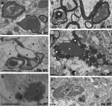
Figure 6.
Ultrastructure of the thalamic degeneration. A: Degenerated myelinated axons display swollen mitochondria and darkly degenerated masses. B: Sections treated with potassium chromate show that mitochondria in degenerating axons contain high calcium levels (white asterisk). C: Dark degenerated corticothalamic axon terminals maintain synaptic contact zones with dendrites (d); a, astroglial process. D: Degenerated neuron with dense nucleus (n) and dense cytoplasm contains swollen mitochondria, partly staining for calcium deposition (white asterisk). They are also found in degenerated boutons (b). Section was treated with potassium bichromate. E: Dark degenerated dendrite (d) partially engulfed by astroglia (a) maintains contacts with presynaptic boutons (b). F: Neuronal perikarya contain fibrillar bodies (fb) and accumulations of lysosomes (ly). Scale bar = 0.5 μm.

