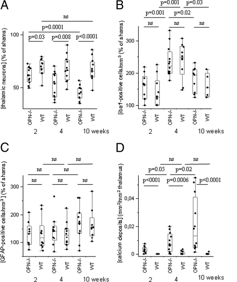
Figure 7.
Morphological and morphometric analyses of the thalamus. A: OPN-KO mice had fewer thalamic neurons than wild-type mice two, four, and ten weeks after the excitotoxic corticostriatal lesion, this loss being progressive only in OPN-KO mice. B: Numbers of microglial cells did not differ significantly between OPN-KO and wild-type mice, and peaked after four weeks survival. C: GFAP-positive cells were more numerous in ibotenate-treated mice compared with sham animals, but their numbers did not differ significantly between ibotenate-treated strains and survival time. D: The total area of thalamic microcalcification was higher in OPN-KO than in wild-type mice and increased further over time.

