Figure 2.
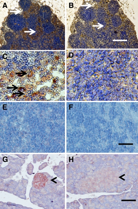
Photomicrographs of mesenteric lymph node (A–F) from a STZ-diabetic rat into which E28 pig pancreatic primordia had been transplanted in the mesentery four weeks previously (A–D) or a nontransplanted nondiabetic rat (E and F) or a pancreas from a nondiabetic nontransplanted rat (G and H) stained using anti-insulin antibody (A, C, E, and G) or control serum (B, D, F, and H). Arrows delineate tissue that stains positive for insulin (red-brown) (A and C) or negative staining tissue (B). Arrowheads delineate islet of Langerhans (G and H). Scale bars: 80 μm (A and B); 30 μm (C–F); 100 μm (G and H).

