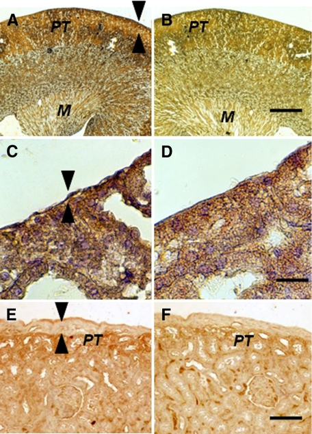
Figure 6.
Photomicrographs of the contralateral kidney from a diabetic rat into which embryonic pig pancreas had been transplanted in the mesentery and pig islets had been implanted subsequently in the other kidney (A–D) or of a kidney from a diabetic rat in which pig islets had been implanted without prior transplantation of E28 pig pancreatic primordia (E and F) stained using anti-insulin antibody (A, C, and E) or control antiserum (B, D, and F). Arrowheads delineate a normal sized subcapsular space (A, C) or expanded subcapsular space (E). PT, A, B, E, and F. M, medulla (A and B). Scale bars: 100 μm (A and B), 10 μm (C and D), or 40 μm (E and F).

