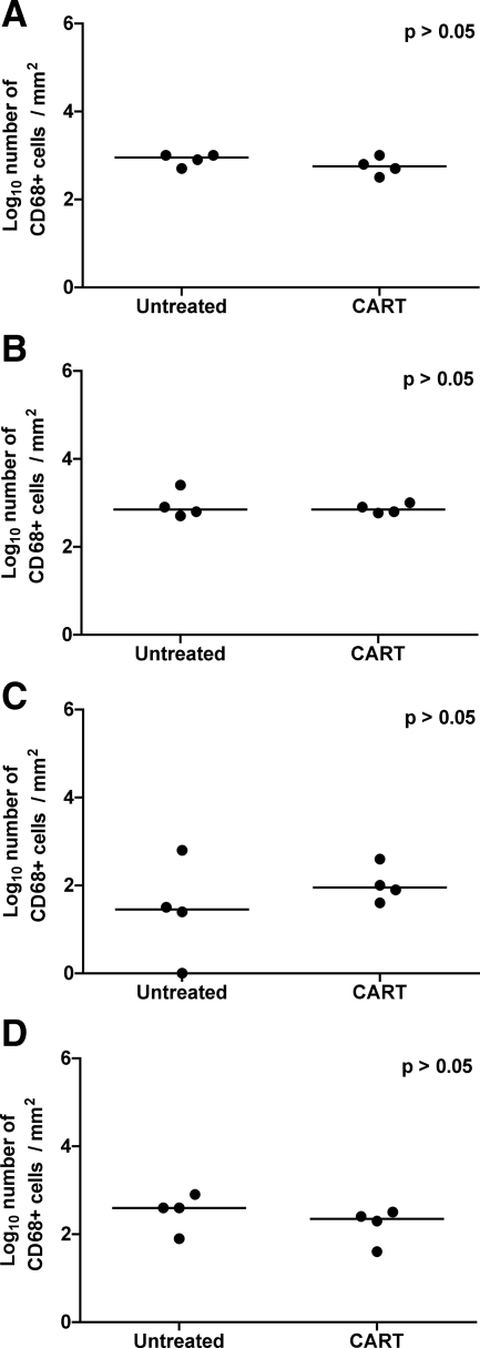
Figure 9.
Immunohistochemistry for CD68 in brain. Quantitative immunohistochemistry for CD68 in frontal cortex (A), brainstem (B), hippocampus (C), and putamen (D) from untreated controls and macaques that received CART, reported as positive particles/mm2 of tissue. Note that significant differences were not observed in any of the regions of brain. Horizontal bars indicate group median values.

