Figure 5.

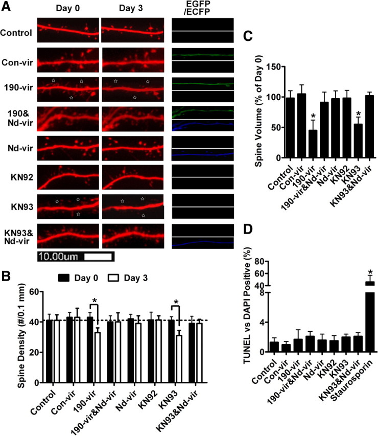
NeuroD activity is required to maintain spine stability. Primary cultures were treated with PBS (control), con-vir, 190-vir, 190-vir plus nd-vir, nd-vir, KN92 (2 μm), KN93 (2 μm), or KN93 (2 μm) plus nd-vir for 3 d. A, Live-cell images of a portion of a single dendrite from a representative neuron on day 0 and day 3. EGFP and ECFP were used to indicate successful infection of con-vir/190-vir and nd-vir, respectively. Stars indicate the spines' decrease in size. B, Spine densities on day 0 and day 3 were quantified. C, Spine volumes were calculated by the overall DsRed fluorescence and the fluorescence intensity on day 3 was normalized against that on day 0. D, he apoptosis rates were also determined, with neurons treated with 1 μm staurosporin for 24 h serving as positive control. Error bars indicate SD. *p < 0.05.
