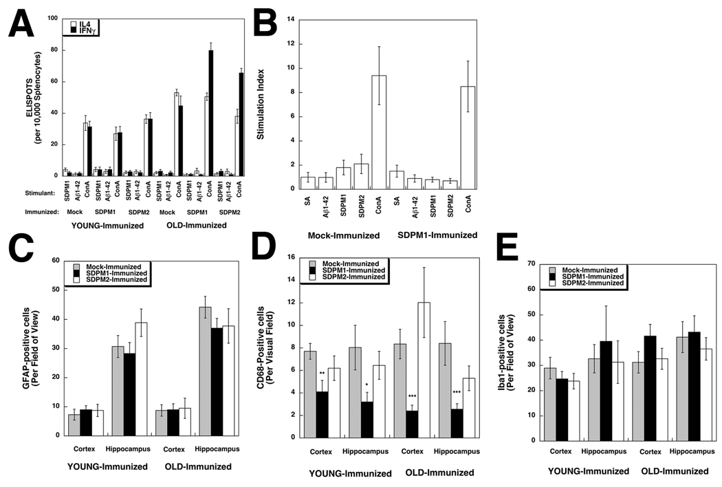
Figure 5. Immunization with SDPM1 does not stimulate T cell responses to SDPM1 or Aβ1–42 or induce brain inflammation in APPswePSEN1(A246E) transgenic mice.
(A) Quantification of ELISPOT assays for Interleukin 4 (IL4)-positive and Interferon gamma (IFNγ)-positive T cells after stimulation with SDPM1, Aβ1–42 peptide, or Concanavalin A (ConA), subtracted from no peptide control. (B) Splenocytes were stimulated with streptavidin (SA), Aβ1–42, SDPM1, SPDM2, or Concanavalin A (ConA), after which cells were incubated with 3H thymidine. 3H incorporation was assayed to determine stimulation of cell division of T lymphocytes, relative to serum-containing media alone. Cells staining positive for GFAP (C), CD68 (D), or Iba1 (E) were quantified. Errors are SEM for n=7–12 animals per condition for n=3 measurements per animal in (A), 4 measurements per animal in (B), and 10 measurements per animal in C–E.

