Figure 1.
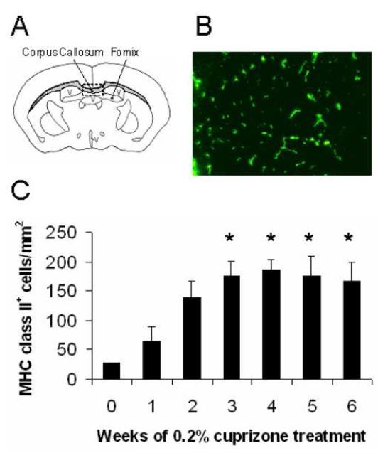
MHC class II+ cells were detected in the corpus callosum of cuprizone-treated mice.
(A) Illustration of the corpus callosum from which all coronally-sectioned mouse brains were analyzed near midline within the rectangular box. V = ventricle
(B) Brain sections were stained with anti-I-Ab antibody to identify MHC class II+ cells in the demyelinated corpus callosum at week 3.0 (200X magnification).
(C) MHC class II+ cells were quantified at weekly intervals during the 6 week exposure to cuprizone and were compared to untreated mouse brains (week 0). *Denotes p<0.05 by one-way analysis of variance (ANOVA) test followed by Dunnett multiple comparisons test comparing all time points to 0 week timepoint; n=3 mice each week

