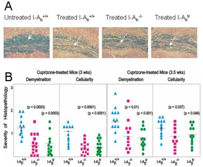
Figure 2.
Mice exhibited reduced demyelination in the corpus callosum when MHC class II was absent or when the cytoplasmic tail was truncated.
(A) LFB-PAS-stained coronal brain sections exposing the corpus callosum (indicated by the arrow) showed blue-stained myelin in untreated I-Aβ+/+ mice. Treatment of wild type, I-Aβ−/−, and I-Aβtr mice with cuprizone for three weeks induced demyelination that is indicated by the lack of blue and the presence of pink counterstained fibers. Wildtype mice had significantly greater demyelination (indicated by the decreased blue staining) compared to I-Aβ−/− and I-Aβtr mice. All pictures are shown at 100X magnification.
(B) The histopathology of the corpus callosum in the I-Aβ−/− and the I-Aβtr mouse was significantly reduced when compared to the wild type mouse. Brain sections were scored by three independent readers in a double-blind manner for the severity of demyelination and cellularity on a scale of zero to three and averaged as previously described (Hiremath et al., 1998). A score of 3 indicated complete demyelination, and a score of 1.5 indicated demyelination of 50% of the fibers. A score of zero indicated the lack of demyelination. Similarly, peak cellularity indicated by numbers of nuclei at week 5 of cuprizone treatment was taken as a score of 3 while untreated control was given a score of zero. Statistical analyses were conducted by ANOVA followed by the Dunnett multiple comparisons test which compared the wild type to I-Aβ−/− or I-Aβtr mice. Results are from three independent experiments using 4 mice per group per time point.

