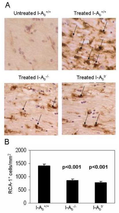
Figure 3.
The absence of MHC class II or the cytoplasmic tail of MHC class II showed a reduced microglia/macrophage accumulation in the corpus callosum.
(A) Representative RCA-1-stained microglia/macrophages (brown-stained cells with nuclei and indicated by arrows) in the corpus callosum of untreated controls wild type, I-Aβ−/−, and I-Aβtr mice treated for 3 weeks with 0.2% cuprizone at 400X magnification are shown.
(B) Morphometric quantitation of RCA-1+ microglia/macrophages were facilitated by using the NIH Imaging software (Hiremath et al., 1998). The corpus callosum of wild type mice treated with cuprizone (n = 12 per genotype) for three weeks had greater numbers of microglia/macrophages than I-Aβ−/− or I-Aβtr mice. Statistical analyses were conducted by ANOVA using the Dunnett multiple comparison test comparing to wild type mice.

