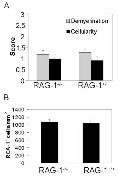
Figure 7.
Cuprizone-induced neuropathology and microglia/macrophage numbers were not affected by the RAG-1−/− mutation.
(A) RAG-1−/− and RAG-1+/+ mice exhibit a similar degree of demyelination and cellularity at 3 weeks (n = 11-12 per genotype). Three independent experiments were conducted using 3 or 4 mice per genotype. LFB-PAS- and RCA-1-stained coronal brain sections were scored as described in Figure 2 and Figure 3, respectively.
(B) Quantitation of the RCA-1+ microglia/macrophages revealed no statistical difference in the brains of RAG-1−/− and RAG-1+/+ mice (n = 11-12 per genotype) as described in Methods section. Statistical analysis was conducted using ANOVA comparing wild type to RAG-1−/− mice.

