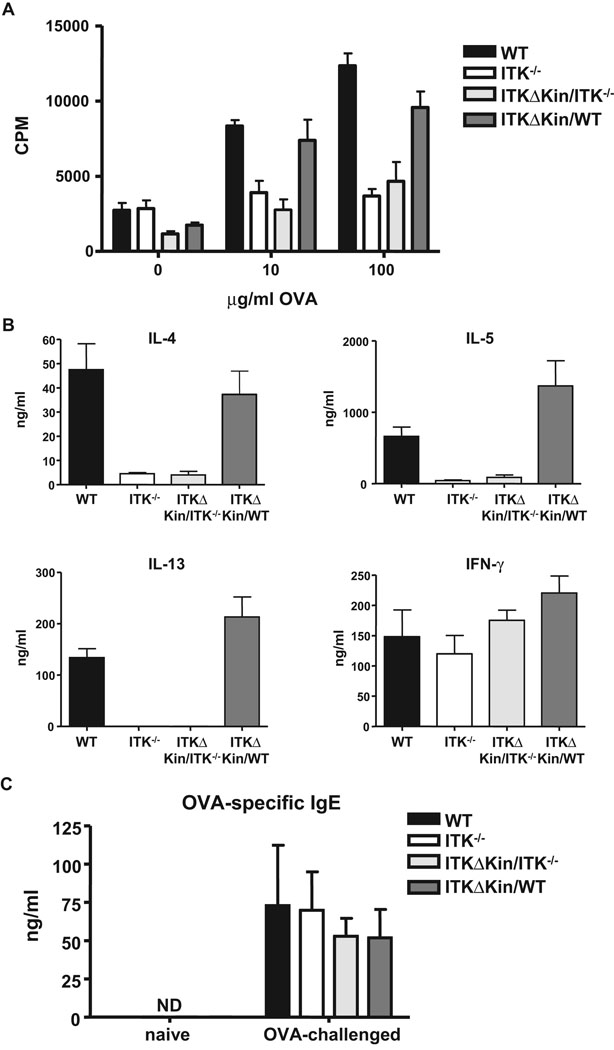
Figure 3. Role of the kinase domain of Itk in T cell responses and Th2 development in response to OVA immunization.
(a) WT, Itk−/−, Tg(Lckpr-ItkΔKin)/Itk−/−, and Tg(Lckpr-ItkΔKin)/WT mice were treated as in figure 1, and spleens and lymph nodes collected, stimulated with the indicated concentration of OVA, and proliferation determined. (b) Supernatants from splenocytes and lymph node cells stimulated with 100 µg/ml OVA in vitro for 96 hours, were collected and assayed for IL-4, -5, -13 and IFNγ. (c) WT, Itk−/−, Tg(Lckpr-ItkΔKin)/Itk−/−, and Tg(Lckpr-ItkΔKin)/WT mice were treated as in figure 1, and serum collected and analyzed for OVA specific IgE as described in the materials and methods section. *p<0.05 vs. WT mice; ND, none detected.

