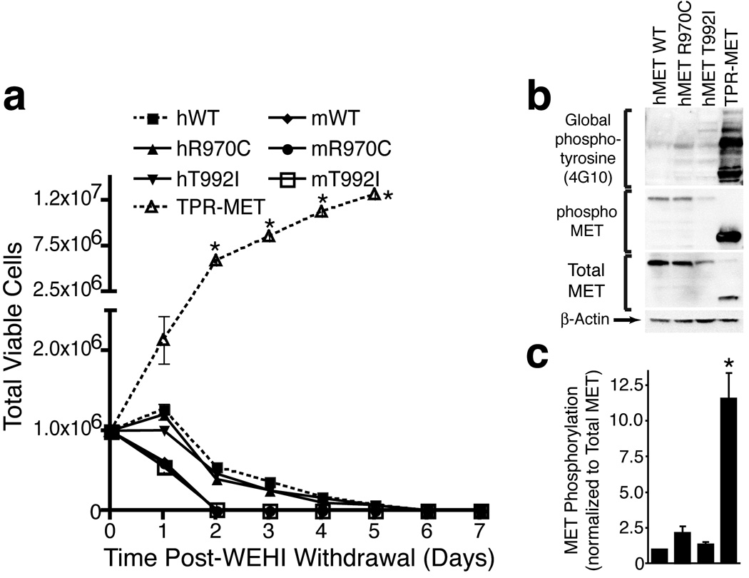
Figure 1. Characterization of MET Sequence Variants in Ba/F3 Cells.
(A) Ba/F3 cells overexpressing human or murine METWT, human METR970C, human METT992I, murine MetR968C, murine MetT990I, or TPR-MET were plated in media without IL-3. Cells were counted daily for one week. Values represent mean ± s.e.m. (n=3). * indicates p < 0.002 with t-test in comparison to human METWT.
(B) Whole cell lysates from 293 T17 cells overexpressing human METWT, METR970C, METT992I, or TPR-MET were subjected to immunoblot analysis with antibodies specific for phospho-tyrosine (4G10), phoshpo-MET, total MET, or β-actin.
(C) Densitometric analysis of immunoblots shown in panel (B). Phospho-MET is normalized to total MET and values are expressed as fold of WT. Values represent mean ± s.e.m. (n=3). * indicates p < 0.05 with t-test in comparison to human METWT.

