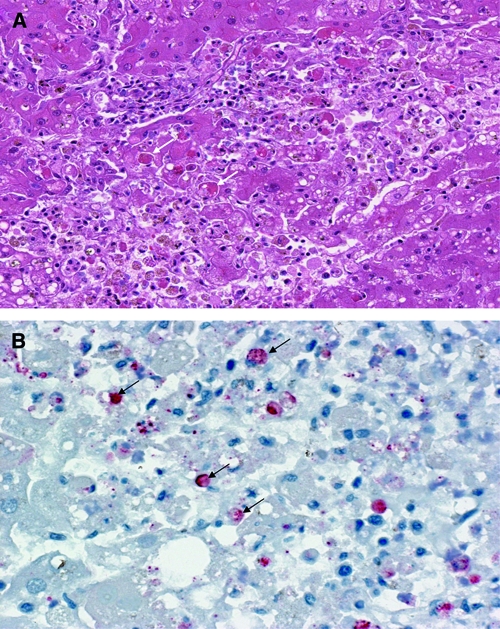
Figure 4.

(A) Histopathological and (B) immunohistochemical analyses of the liver tissues from Rift Valley fever (RVF) cases during the outbreak in Tanzania, 2007. (A) Shows liver section with hematoxylin and eosin stain revealing extensive hepatocellular necrosis with acidophilic material in the cytoplasm (arrows). (B) Shows liver section with immunohistochemical staining for RVF viral antigens showing positive immunoreactivity in hepatocytes and Kupffer cells (arrows).
