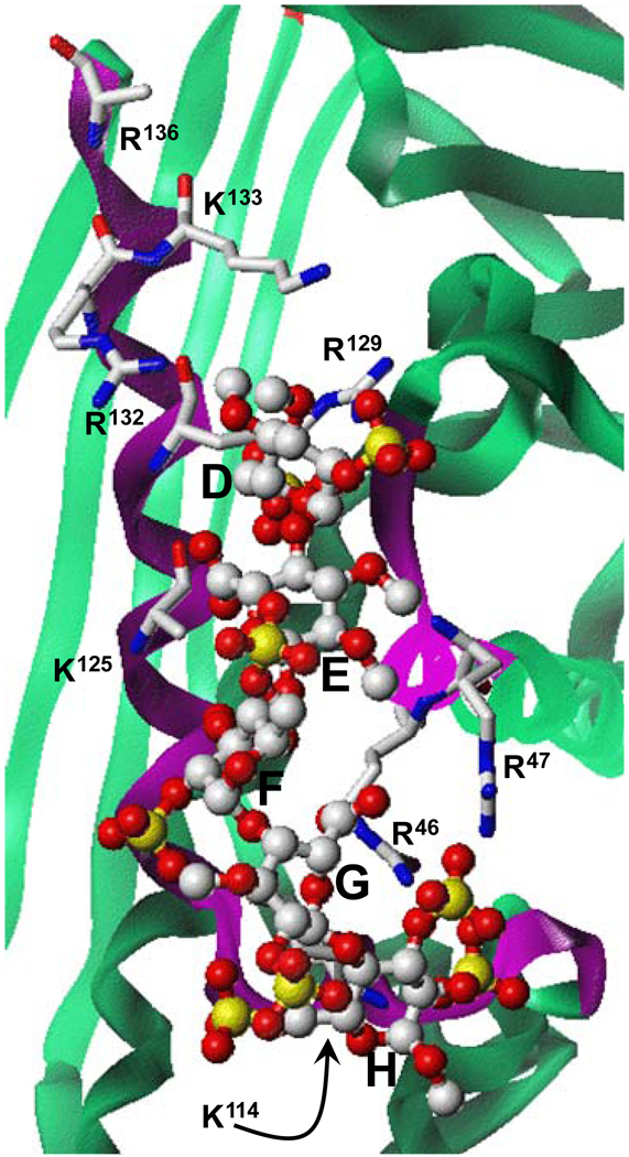
Figure 3. Close-up view of the heparin-binding site in antithrombin.
The structure of co-complex was obtained from PDB (filename ‘1e03’). Green ribbon shows antithrombin and magenta represents the heparin-binding site. Pentasaccharide DEFGH is shown in ball-and-stick representation. Extensive interactions between antithrombin arginine and lysines with multiple sulfate groups of DEFGH engineer the high affinity, high specificity interaction. Majority of the non-ionic binding energy involved in the heparin – antithrombin interaction is thought to arise from the hydrogen bond type interaction with sulfate groups. Figure modified from Desai UR. Med. Res. Rev. 2004; 24:151–181).

