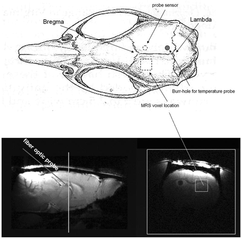
Figure 1.

Temperature probe sensor, burr hole and MRS voxel locations. The upper image is a diagram of rat skull (dorsal view) with the indicated skull land marks: Lambda and Bregma (Paxinos and Watson, The Rat Brain in Stereotaxic Coordinates. Second Edition. New York: Academic Press, 1986). The bottom panels are sagital (left) and transaxial (right) images from a rat brain (note the transaxial image is from the plane crossing the white line indicated in the sagital image). The temperature probe was inserted from burr-hole through the skull at a 45° angle and the probe sensor reached the region that neighbored the MRS voxel (dashed circle in the upper sketch, and dark region in the bottom right transaxial brain MR image). The voxel size is 3 × 3 × 3 mm3 and positioned as indicated.
