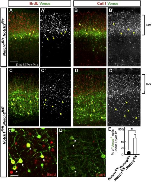
Figure 3. Notch deletion causes laminar displacement of later-born neurons.
(A-D”) Venus (green) immunostaining with BrdU or Cutl1 (red or white) staining in indicated mutants at P14. Note that Cutl1 analysis was performed in the dorsal somatosensory region where endogenous Cutl1 expression in lower layers is normally absent. BrdU+/Venus+ or Cutl1+/Venus+ neurons in the Notch deleted cortex (C-D’, arrows) are abnormally positioned in lower layers than in the control cortex (A-B’, arrows). (C”, D”) Higher magnification views of BrdU+/Venus+ and Cutl1+/Venus+ double-labeled neurons around layer V, respectively (arrows). Bars (μm) = 100 (A-D”), 10 (C”, D”’). (E) Quantification of the distribution of Venus+ neurons under Layer IV in the Notch1 fl/+; Notch2 fl/+ and Notch1 fl/fl; Notch2 fl/fl cortex. The data represent the mean ± SEM of 4 brains each from independent experiments. * p<0.005, Student’s t-test.

