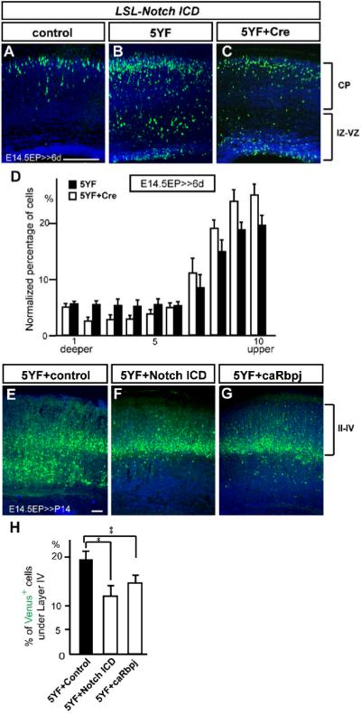
Figure 7. Notch ICD mitigates radial migration defects induced by a dominant-negative form of Dab1.
(A-C) Venus+ neurons were detected by immunohistochemistry 6 days after electroporation with pCDNA3.1 empty (A) or containing Dab1 5YF (B, C) plasmid with pTα1-IRES-Venus (A, B) or pTα1-Cre-IRES-Venus (C) plasmid in LSL-Notch ICD mice. Note that 5YF arrested many neurons beneath the CP which was mitigated by overexpression of Notch ICD. (D) Quantification of neuronal distribution in the LSL-Notch ICD cortex electroporated with indicated plasmids (K-S test, p<0.05; Repeated Measures ANOVA, F(9,126)=5.24, p<0.05). The data represent the mean ± SEM of 6 brains each. (E-G) Venus+ neurons were detected by immunohistochemistry at P14 after E14.5 electroporation with pCALSL empty (E), containing Notch ICD (F) or caRbpj (G) with pTα1 -Cre-IRES-Venus and pCDNA3.1-Dab1 5YF plasmids in wild-type mice. Note that compared to control (E), overexpression of Notch ICD (F) or caRbpj (G) resulted in fewer neurons located beneath layer IV. (H) Percentages of Venus+ neurons below layer IV in P14 wild-type cortex electroporated with indicated plasmids. The data represent the mean ± SEM of 10, 8, and 11 brains, respectively. *,**p<0.01, 0.05, by Student’s t-test. Bars = 100 μm.

