
FIGURE 4. Microglial cell staining.
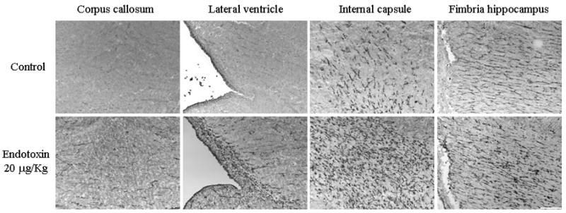
Brain sections from post-natal day 1 control and endotoxin rabbits were stained for microglial cells using tomato lectin. An increase in activated microglial cells is noted in the endotoxin treated kits when compared to the control kits in the regions of the corpus callosum, along the borders of the lateral ventricle, region of the internal capsule and the fimbria hippocampus. Scale bar is 200 μm.