Figure 1.
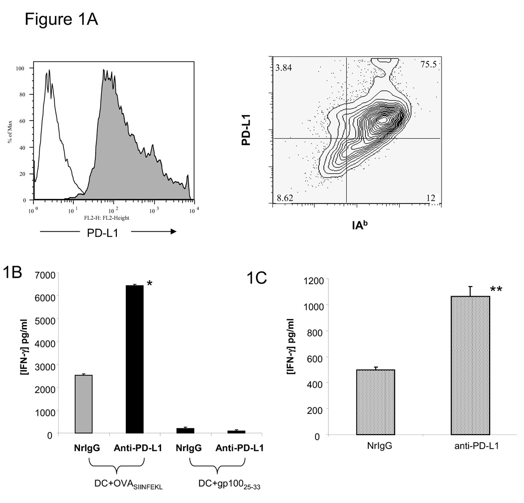
Peptide-loaded DC pre-treated with anti-PD-L1 antibody enhance stimulation of a specific T cell response. (A) After 5 days of in vitro culture, bone marrow-derived DC were stained with anti-IAb and PD-L1 antibodies and analyzed by flow cytometry. Filled histogram = anti-PD-L1, open histogram = isotype control. (B) DC were pulsed with OVA257–264 or gp10025–33 peptide and treated with either 10 µg/ml NrIgG or anti-PD-L1 antibody for 24 hours. These DC were then co-cultured with OT-I splenocytes at a 1:10 ratio for 48 hours. (C) DC were pulsed with OVA323–337 peptide and treated with either 10 µg/ml NrIgG or anti-PD-L1 antibody for 24 hours. These DC were co-cultured with OT-II splenocytes at a 1:10 ratio for 48 hours. Supernatants were collected and tested in an IFN-gamma ELISA assay. One of 2 representative experiments is shown. *indicates p<0.001, **indicates p<0.01.

