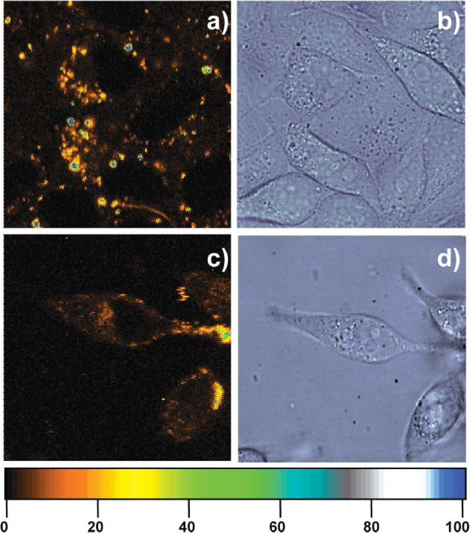
Fig. 2.
(a) Confocal fluorescence and (b) corresponding DIC images of HeLa cells incubated for 24 h with 1 µL of the Lipofectamine/C24:Agn complexes at 37 °C under 5% CO2. [C24] = 0.15 µM, [Ag+] = 1.8 µM, and [BH4−] = 1.8 µM. (c) Confocal fluorescence and (d) corresponding DIC images of HeLa cells incubated for 24 h with 20 µL of the C24:Agn at 37 °C under 5% CO2. [C24] = 3 µM, [Ag+] = 36 µM, and [BH4−] = 36 µM. The living cells were taken out of the incubator after 24 h and confocal fluorescence images were obtained using a scanning stage (3 ms exposure per pixel, excitation intensity 130 W cm−2). The colour scale bar indicates the emission rate of the confocal fluorescence microscopy images in counts per ms.

