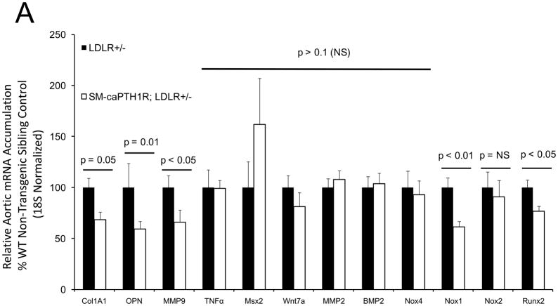
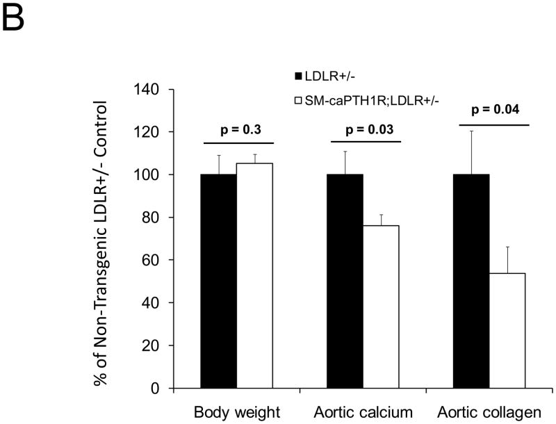
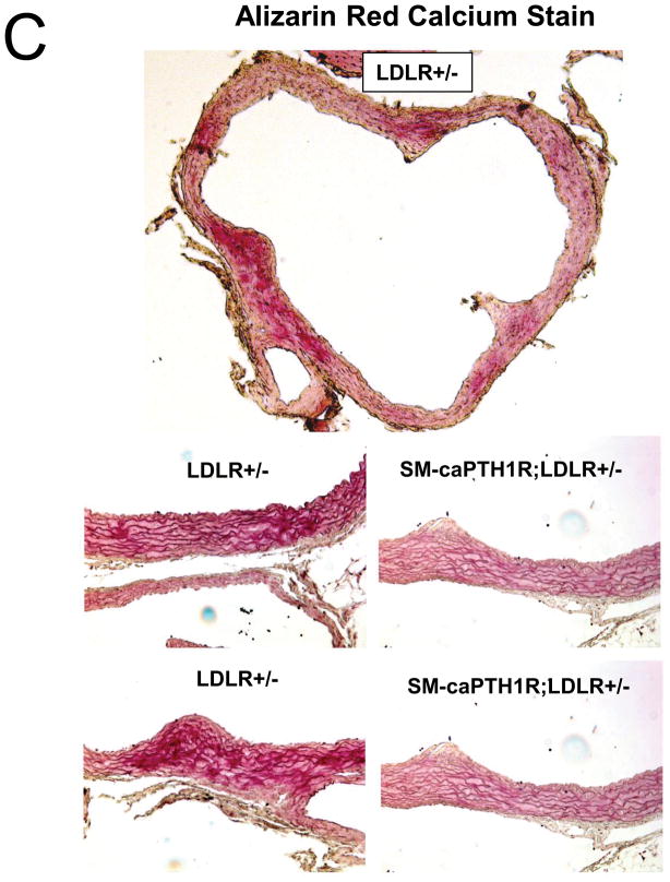
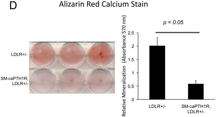
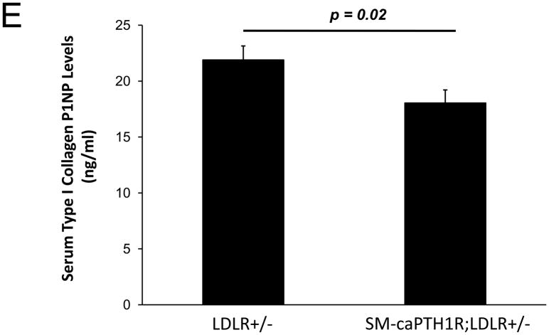
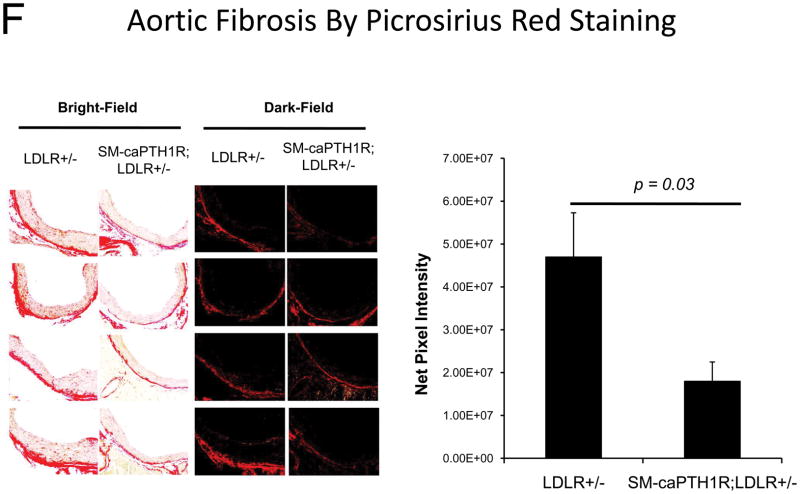
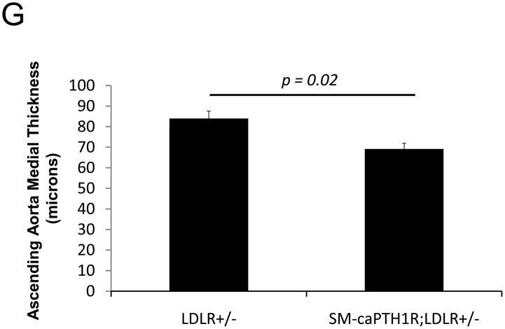
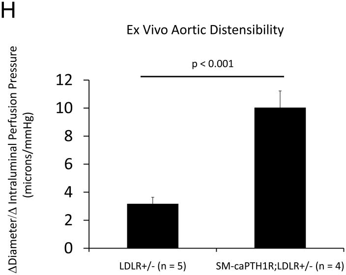
Figure 5. The SM-caPTH1R transgene down-regulates aortic fibrosis and calcification without inhibiting TNF, BMP2, Wnt, and Msx2 gene expression in diabetic LDLR+/− mice.
Panel A, aortic Col1A1, OPN, MMP9, Nox1, and Runx2 were reduced in LDLR+/− mice possessing the SM-caPTH1R transgene. Nox4 and upstream osteogenic signals were not altered (n = 10; 4 months HFD). Panel B, aortic calcium and collagen content were decreased in aortic extracts from diabetic LDLR+/− mice possessing the SM-caPTH1R transgene (n = 6–12 per group; 3 months HFD). Panel C, aortic calcium deposition occurred primarily within the tunica media. Panel D, the SM-caPTH1R transgene suppressed VSMC calcification in vitro. Panel E, serum P1NP, a marker of collagen biosynthesis, was also decreased (n = 19–22 per group). Panel F, aortic collagen content assessed by Picrosirius histochemistry and digital image analysis confirmed that SM-caPTH1R transgene decreased fibrosis (n = 4). Panel G, thickness of the aortic tunica media was reduced by the SM-caPTH1R transgene. Panel H, aortic distensibility was increased by the SM-caPTH1R transgene.

