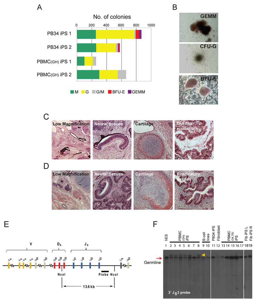
Figure 2. Pluripotency and V(D)J re-arrangement of peripheral blood-derived iPS cells.
(A) Embryoid bodies derived from PB34 and PBMC iPS cells yield hematopoetic colonies in semisolid methylcellulose media: burst forming unit-erythroid (BFU-E), colony forming unit-granulocyte (CFU-G), colony forming unit-macrophage (CFU-M), colony forming unit-granulocyte, macrophage (CFU-GM) and colony forming unit-granulocyte, erythroid, macrophage (CFU-GEMM). Total number of each type of colony was counted.
(B) Representative images of various types of hematopoietic colonies. Images were acquired with a standard microscope (Nikon, Japan) with a 20x objective.
(C, D) Hematoxylin and eosin staining of teratomas derived from immunodeficient mice injected with PB34 iPS (C) and PBMC iPS (D) cells show tissues representing all three embryonic germ layers.
(E) Genomic DNA from peripheral blood-derived iPS lines grown was digested with Nco I and analyzed for V(D)J rearrangements at the TCRδ (T-cell receptor Delta) locus by Southern blotting using a 3’Jδ3 probe.
(F) TCRδ V(D)J recombination of blood-derived iPS cell clones. Lanes 2–8 and lanes 13–17 are PBMC iPS lines. B-cell lines on lanes 8 and 9 showed no rearrangement. TCRδ rearrangement was observed for some PBMCs derived iPS lines (lanes 2–6 and 8). Lanes 1, 11, 12, 18, 19 are H1 hES cells, PB34 iPS cells, fibroblast cells, fibroblast derived iPS using retrovirus and lentivirus, respectively. The red arrow indicates expected size of the germline band. Orange arrow indicates re-arranged bands.
For further information on the pluripotency, V(D)J rearrangement and fingerprint analysis performed on the peripheral blood-derived iPSC clones see also Figures S2 and Table S2.

