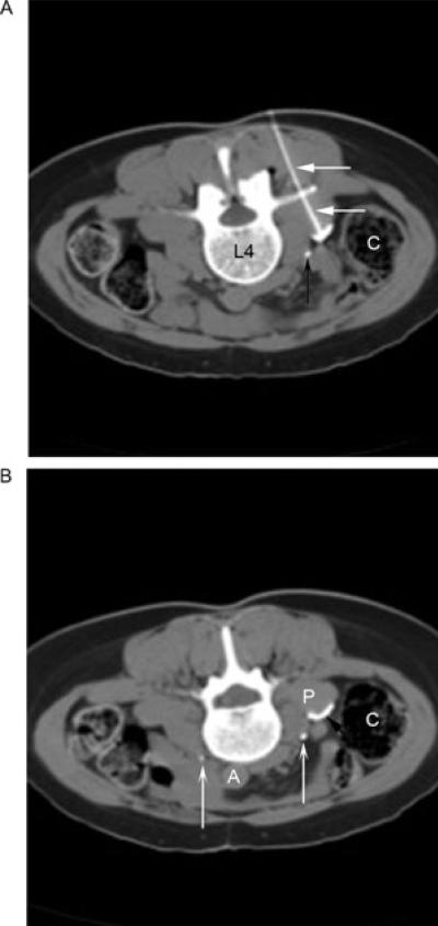
Figure 1.
(A) An axial computed tomography (CT) image in soft tissue window with the patient in prone position demonstrates the needle (straight white arrows) being advanced under CT and stimulating guidance until sensory stimulation at 50 Hz was achieved at 0.35 V. Concave black arrow, right ureter. C = ascending colon; L4 = fourth lumbar vertebral body. (B) A later axial CT image in soft tissue window demonstrates 0.5 mL of Omnipaque with phenol spreading (straight black arrow) on the anterior surface of the psoas muscle (P), but remaining medial and posterior to the ureter (concave white arrows, right and left ureters) and lumbar sympathetic chain, posterior and medial to the colon (C), and anterior to the lumbar nerve roots. A = aorta.

