
Figure 3.
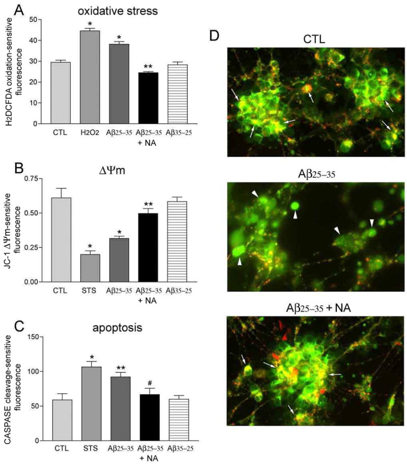
NA inhibits Aβ-induced oxidative stress, mitochondrial depolarization, and caspase activation. A) Bar graph shows that 1 hour of 10 μM Aβ25–35 results in increased reactive oxygen species (ROS) in hNT neurons. 10 μM NA restored ROS to CTL levels. Hydrogen peroxide (H2O2) was used as a positive control. No effect was seen with reverse Aβ. *, p < 0.001 vs. CTL; **, p < 0.001 vs. Aβ. B) NA prevented depolarization of the hNT mitochondrial membrane potential (Δψm) after 24 hours of treatment with Aβ. Δψm was measured using a JC-1-dependent ratio of intact (red/green) to depolarized (green) mitochondria. Staurosporine (STS) was the positive control. *, p < 0.001 vs. CTL; **, p < 0.05 vs. Aβ. C) 24 hours of Aβ exposure increases caspase activation, which was inhibited by NA. *, p < 0.001 vs. CTL; **, p < 0.05 vs. CTL; #, p < 0.05 vs. Aβ; n = 8/treatment group in 3 independent experiments for each assay. D) Fluorescent photomicrographs show overlays of intramitochondrial JC-1 aggregates (red) and intracellular JC-1 (green) in hNT cultures treated 24 hours in CTL (top), Aβ (middle), or Aβ with NA (bottom) conditions. Arrows in top and bottom panels show distinct red + green JC-1 colocalization indicating healthy mitochondria. Arrowheads in middle panel show diffuse green JC-1 staining of depolarized mitochondria. Values in the Y-axis for (A–C) are arbitrary spectrophotometric units.