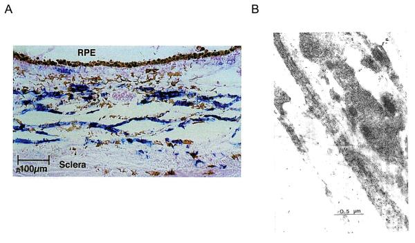
Figure 7.
Non vascular smooth muscle of the primate (A) and chick (B) eyes. A. Suprachoroid and inner sclera contain cells positive for smooth muscle α-actin (blue chromogen counterstained with nuclear fast red). Reproduced with permission from Poukens et al., 1998 © Association for Research in Vision and Ophthalmology. B. Electron micrograph of choroidal cells labeled with antibodies to smooth muscle actin (black dots), showing long fibers not associated with blood vessels. M, muscle cell; F, fibroblast; C, collagen. Reproduced with permission from Wallman et al., 1995 © Association for Research in Vision and Ophthalmology.

