Fig. 3.
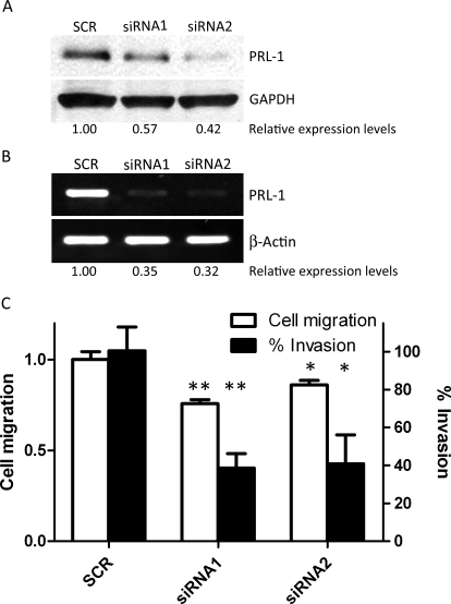
Depletion of PRL-1 inhibits cell migration and invasion. A, Western blotting detects PRL-1 protein levels at 24 h after siRNA transfection. Protein expression levels are relative to GAPDH. B, reverse transcription-PCR detection of PRL-1 mRNA levels 24 h after siRNA transfection in A549 cells. mRNA levels are relative to β-actin. C, PRL-1 siRNA inhibits cell migration in the scratch wound-healing assay. Transiently siRNA transfected A549 cell monolayers were disrupted with a sterile micropipette tip. The number of cells in the denuded zone was determined at the indicated times (0 or 72 h) by inverted microscopy. Quantitative assessment of the mean number of cells in the denuded zone is expressed as mean ± S.D. The experiment was repeated three times. ∗, P < 0.05; ∗∗, P < 0.01, compared with scrambled control. B, PRL-1 siRNA inhibits cell invasion and migration. Cell invasion of transiently siRNA transfected A549 cells was assessed at 24 h using Matrigel invasion chambers, as described under Materials and Methods. Three fields in each well were counted, and the mean percent invasion through the Matrigel matrix membrane was determined relative to the migration through the control membrane. The bar graph presents the mean relative values obtained from three independent determinations (±S.D.). ∗, P < 0.05; ∗∗, P < 0.01, compared with the scrambled control.

