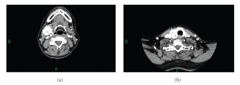
Figure 1.
CT of neck with contrast. (a) Well-defined tumour mass in the right submandibular gland measuring 29 × 26 × 30 mm. It shows prominent vascularisation which would be unusual for benign lesion such as PA. There is some low-density area centrally in the tumour probably related to necrosis. (b) There is a large nodule in the thyroid on the right side measuring 26 × 23 × 30 mm showing similar intense enhancement as the submandibular lesion.

