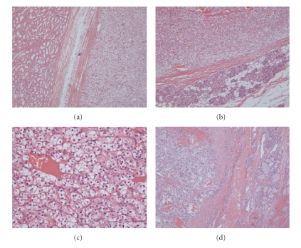
Figure 2.
(a) Original renal tumour (H&E stain, original magnification x40). On the left is normal kidney and clear cell RCC is on the right. (b) Submandibular gland (H&E stain, original magnification x40): metastatic RCC on the top with normal glandular tissue in the lower portion. (c) Metastatic RCC in submandibular gland (H&E stain, original magnification x200). (d) Thyroid tumour (H&E stain, original magnification x40): tumour on the left and normal thyroid tissue on the right.

