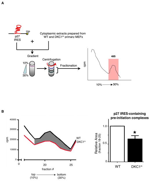
Figure 2. Molecular mechanism of p27 IRES-mediated translation impairment in DKC1m cells.
A) Schematic of the biochemical approach used to study the 48S complex formation in WT and DKC1m MEFs. A [32P]-p27 IRES mRNA probe was incubated with cytoplasmic extracts. Newly assembled 48S complexes were separated by sucrose gradient centrifugation and fractionated according to their density. A peak of radioactivity was generated and coincided with fractions containing the 48S complexes. B) Representative of the 48S subunit quantification from total cytoplasmic extracts prepared from serum starved (0.1% FBS) WT and DKC1m cells. Representative profile of a sucrose density gradient reporting the radioactive intensity per fractions in WT (black) and DKC1m (red) extracts, respectively (left). Columns, mean ± SEM of the area under the curve in WT and DKC1m cell extracts measured in 3 independent experiments; bars, SEM (right). Statistical analysis was carried out using Student t test. *P<0.05.

