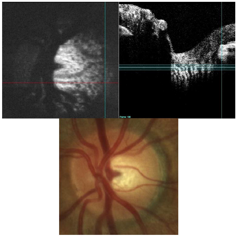
Fig. 11.

Pores in the lamina cribrosa (black spots, upper left) are clearly visible in a normal subject with a large disc (upper left). Note the enhanced visualization on the temporal side of the disc compared to the clinical view provided by the fundus photo on bottom. The C-mode slab is located in the lamina cribrosa/pre-lamina region of the optic nerve (upper right). The 3D dataset is presented as a C-mode sequence (View 3). The upper portion of this figure is one of the slices from the 3D dataset, with the stack of C-mode slices on the left, and the single frame repeated on the right.
