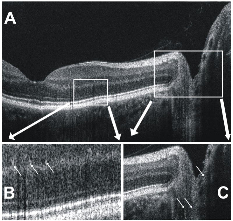
Fig 2.

(A) A closer look at the SdOCT B-scan image in Fig. 1 shows (B) the visualization of small white granular-appearing structures (arrows) in the inner and outer plexiform layers of the retina. (C) Similarly, faint structures appear (arrows) to be visualized in the posterior optic nerve head.
