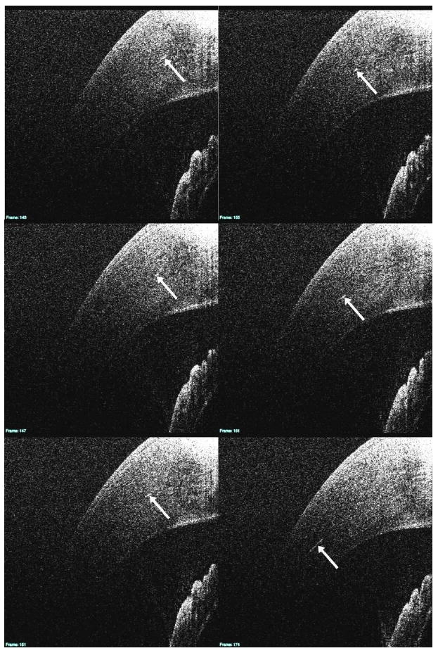
Fig. 7.

In this series of cross-sectional B-scan images, a number of hyper-reflective structures within the stroma are visible (white arrows). The 3D B-scan sequence is linked (View 1). In addition to the slices seen above, it also contains an en-face view of the cornea to the left of the slices.
