
Figure 4.
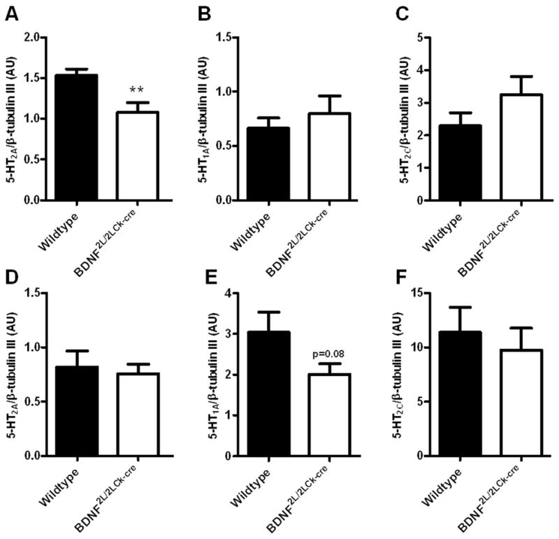
RT-qPCR on 5-HT2A (A, D), 5-HT1A (B, E) and 5-HT2C (C, F) mRNA in frontal cortex (upper panel) and hippocampus (lower panel) of wildtype and BDNF2L/2LCk-cre mice. There was a significant lower 5-HT2A mRNA expression in BDNF2L/2LCk-cre mice in frontal cortex compared to wildtypes (A), while no differences could be observed in hippocampus (D)(n=7–8, Student’s t-test, **P < 0.01). For the 5-HT1A receptor, there was a tendency towards a decrease in expression in BDNF2L/2LCk-cre mice compared to wildtypes (E)(P = 0.08) for hippocampus, while no change was observed in frontal cortex (B). For the 5-HT2C receptor we did not find any differences in expression of mRNA between BDNF2L/2LCk-cre mice and wildtypes. (AU, arbitrary units, Error bars indicate SEM)