
Fig. 5.
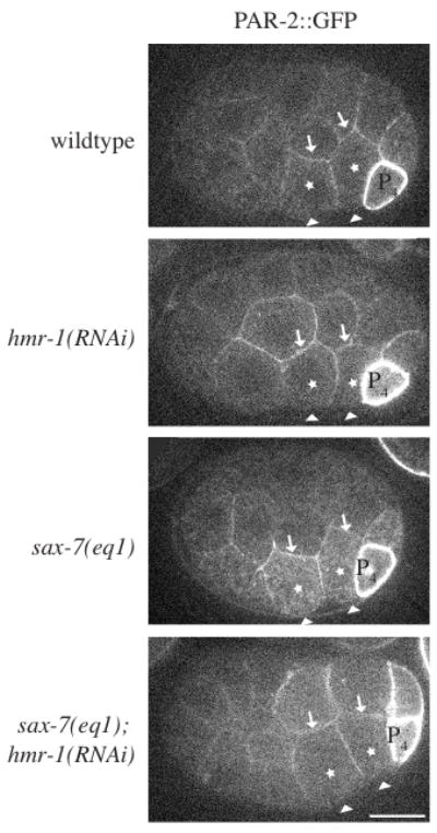
PAR localization indicates Ea/Ep are correctly polarized in sax-7(eq1); hmr-1(RNAi) embryos. PAR-2::GFP was imaged in Ea/Ep shortly following P4 birth in wild-type and mutant embryos. Although GFP distribution was monitored throughout all focal planes, single focal planes are shown. The arrowheads point to the apical surface of Ea/Ep (note that vitelline envelope appears white in sax-7(eq1), but is not the apical surface). Arrows point to the basal surface. The promoter used in the PAR-2 expression construct is highly active in P4 leading to cortical localization. In some embryos PAR-2::GFP is brighter in D than in other cells. This occurs more frequently in sax-7(eq1); hmr-1(RNAi), but has been observed in all genotypes.