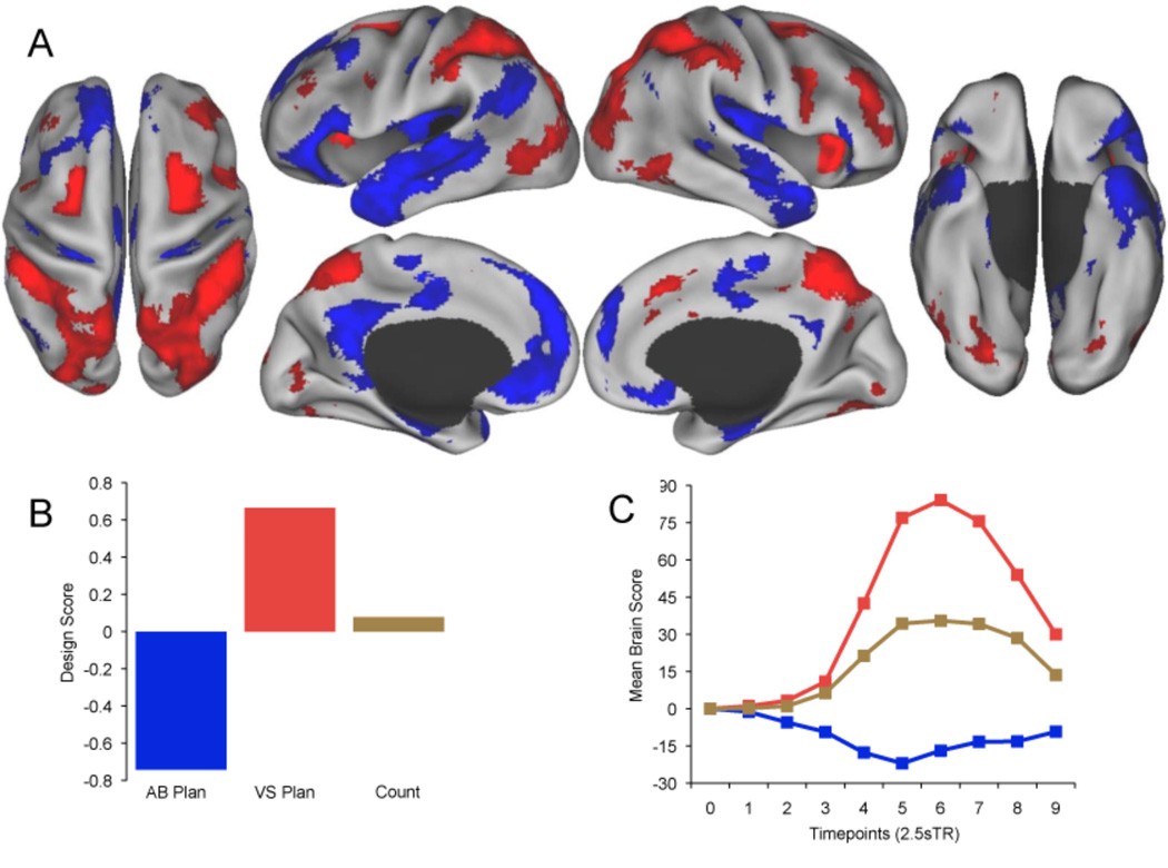
Figure 2.
Task-related brain activity in default and dorsal attention networks dissociated the planning tasks. (A) Activity associated with autobiographical planning (Blue) and visuospatial planning (Red) at TR 6 (17.5 s). Real-time patterns of activation at each TR associated with planning are available as Supplemental Movies 1 and 2. Data are displayed on the dorsal, lateral, medial, and ventral surfaces of the left and right hemispheres of a partially inflated surface map using CARET software (Van Essen, 2005). (B) Design scores for each condition represent the optimal contrast weightings that explain the most task-related variance in BOLD signal. Autobiographical planning was maximally dissociated from visuospatial planning. Counting activity covaried across the same regions as visuospatial planning, although significantly less so. (C) Temporal brain scores convey changes in brain activity related to task at each TR. For each LV, mean brain scores (summary scores of activity across the entire brain of each participant, averaged across participants) show the divergence between experimental conditions over time (Ten 2.5s TRs), and are analogous to hemodynamic response functions typically plotted for individual brain regions.

