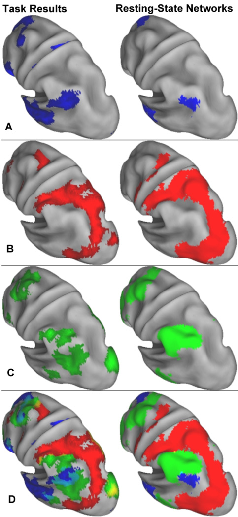
Figure 5.
Lateral parietal view of the PLS and rsfcMRI results. Left parietal lobe activity for autobiographical planning, visuospatial planning and activity common to these two planning tasks (left); and the default, dorsal attention, and frontoparietal control resting-state networks (right). (A) pIPL activity in autobiographical planning subsumes the pIPL cluster in the default resting-state network. (B) Visuospatial planning engaged the same SPL-to-MT+ arc seen in the posterior portion of the dorsal attention network. (C) The two planning tasks commonly engaged a dorsal segment of the aIPL, part of the frontoparietal control network. (D) Overlap of all three networks for task-related activity and resting-state networks. Yellow represents overlap. The cluster ventral to the aIPL in the posterior middle temporal gyrus is suggestive of a concentric ring fitting within the posterior arc of the dorsal attention network. The pIPL region associated with the default network fills this ring. The rsfcMRI results do not mirror this concentric ring pattern, suggesting that it may be task specific. See also Supplemental Fig. 3 for whole brain convergence images of task activity and resting-state network maps.

