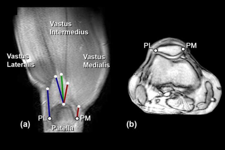
Figure 2.
(a) Anatomic coronal oblique cine-PC image illustrating the corresponding origin and insertion points that define the line-of-action for each musculotendon unit. The imaging plane was aligned parallel to the quadriceps tendon and passed through the myotendinous junction of the vastus intermedius muscle. The myotendinous junctions of the vastus lateralis, vastus intermedius, and vastus medialis muscles can clearly be identified. A similar, but more anterior, imaging plane was used to visualize the mytendinous junction of the rectus femoris (not shown). Moving counter-clockwise in (a): Lines-of-action chosen to represent the vastus medialis muscle are shown in red and represent the distal (VMD) and the proximal (VMP) lines-of-action, respectively. The vastus intermedius line-of-action is shown in green. Lines-of-action for the vastus lateralis muscle are shown in blue and represent the proximal (VLP) and distal (VLD) lines-of-action, respectively. NOTE: Points PM and PL were not chosen from image (a). Rather they were chosen as the most medial and lateral points on the patella from the axial series as illustrated in (b). (b) Mid-patellar axial fastcard image taken at full extension using an imaging plane that bisected the patella in the sagittal plane.

