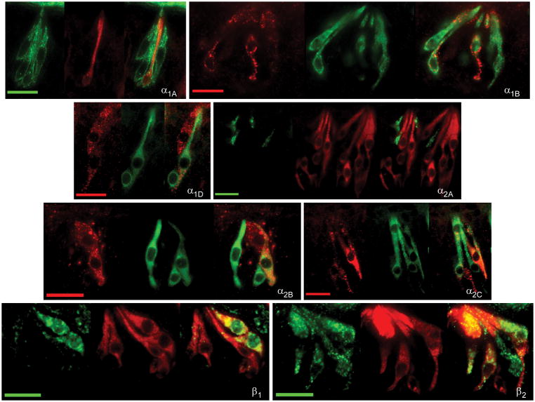
Fig. 4.
Examination of co-expression of adrenergic receptors with gustducin. Representative photomicrographs of double label fluorescent immunocytochemistry with antibodies directed against alpha-gustducin (middle panels) and one of eight tested adrenoceptor subtypes (left panels) are illustrated. Overlaid images are at right of each panel. The scale bar in all photomicrographs represents twenty microns.

