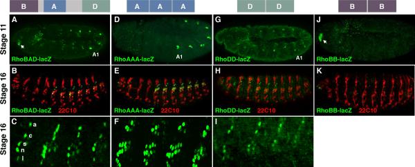
Figure 1. RhoBAD contains separable elements that drive reporter expression in sensory cells.
A-C. Diagram of the RhoBAD enhancer highlighting the relative position of the conserved RhoA, RhoB, and RhoD elements at top. Lateral views of RhoBAD-lacZ stage 11 (A) and stage 16 (B) embryos immuno-stained for β-gal (green) and a PNS-specific neuronal marker (mAb22C10, red) as noted. A lateral view close-up of four abdominal segments shows the β-gal pattern within the lch5 organ (C). The attachment (a), cap (c), scolopale (s), neuron (n), and ligament (l) cells of the C1 lineage are noted. D-F. Diagram of the RhoAAA enhancer at top. Lateral views of RhoAAA-lacZ stage 11 (D) and stage 16 (E) embryos immuno-stained for β-gal (green) and mAb22C10 (red) as noted. A lateral view close-up of four abdominal segments shows the β-gal pattern within the lch5 organ (F). Note, RhoAAA-lacZ is expressed in all the neurons and scolopale cells of the lch5 organ as well as some extra cap and ligament cells. G-I. Diagram of the RhoDD enhancer at top. Lateral views of RhoDD-lacZ stage 11 (G) and stage 16 (H) embryos immuno-stained for β-gal (green) and mAb22C10 (red) as noted. A lateral view close-up of four abdominal segments shows the β-gal pattern within the lch5 organ (I). J-K. Diagram of the RhoBB enhancer at top. Lateral views of RhoBB-lacZ stage 11 (J) and stage 16 (K) embryos immuno-stained for β-gal (green) and mAb22C10 (red) as noted.

