Figure 4.
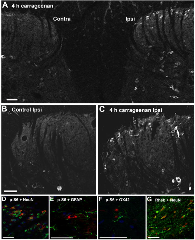
Peripheral inflammation induces phosphorylation of S6 in dorsal horn neurons. (A) Representative confocal microscopy images depicting S6 phosphorylation in the contralateral (contra) and ipsilateral (ipsi) superfical laminae and deep dorsal horn 4 hours after injection of carrageenan to the paw. p-S6 immunoreactivity in the naïve dorsal horn (B) compared to 4 hours after carreageenan injection (C) demonstrates an increase in phospho (p)-S6 following induction of inflammation. The induced p-S6 (red) co-localized with NeuN, a marker for neurons (D), but not with GFAP (E) or OX-42 (F) markers for astrocytes and microglia, respectively. Rheb immunoreactivity colocalized with NeuN (G). DAPI was used as nuclear stain in panels D-F. The scale bar represents 50 μm.

