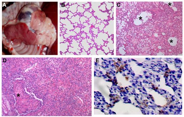
Figure 2. Macroscopic lesions and histological findings of beagles infected with H5N1 virus BHG/QH/3/05.
A) Macroscopic lung lesion of a beagle infected with virus. Day 3 post-intratracheal infection (animal B8). B) A lung section from the control animal that was mock-inoculated with 1 ml PBS i.t. and euthanized on day 5 p. i., HE stain. C) A section from a consolidated area of the lung shows bronchointerstitial pneumonia with significant infiltration of inflammatory cells. The lung lesions were distributed around the bronchioli. Day 3 post-intratracheal infection (HE stain, animal B8). D) Severe alveolar damage within and along the periphery of the consolidated area. Day 5 post-intranasal infection (HE stain, animal B4). Severe proliferative and reactive hyperplasia of alveolar cells with massive recruitment of lymphocytes, fibrin exudates, and alveolar edema are shown. E) Viral antigens in the lung on day 5 post-intranasal infection (brown). IHC. Asterisks indicate lumen of bronchioli (Animal B4).

