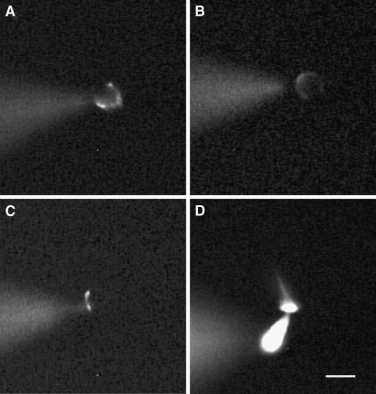
FIG. 1.
Morphology of calyx terminals and type I hair cells as shown by the fluorescent dye Lucifer Yellow, which was included in the patch electrode solution. A–C Images of different calyx terminals surrounding type I hair cells that were loaded with the fluorescent dye during whole-cell recordings. Calyces in A and B appear as horseshoe-shaped structures which surrounded the basolateral regions of the associated hair cell, whereas in C the terminal was more restricted to the base of the type I hair cell. D A type I hair cell loaded with Lucifer Yellow showing the cell body, cuticular plate, and hair bundle. In each image, the dye containing patch electrode is seen to the left of the cell in a different focal plane. Scale bar represents 10 µm.

