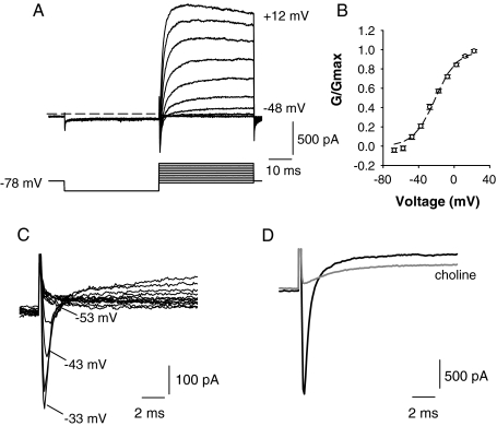
FIG. 3.
A Typical whole-cell currents (upper) recorded from a calyx terminal in extracellular L-15 solution. Voltage protocol is shown in the lower panel. The cell was held at −78 mV and given a 40-ms hyperpolarizing pulse to −128 mV before stepping to test potentials in 10-mV increments between −88 and 12 mV. Dashed line indicates zero-current potential. B Plot showing activation of the peak outward current (G/Gmax) as a function of membrane potential for nine cells (mean ± SEM). A single Boltzmann fit to the data gave a half-activation of −21.3 ± 1.8 mV and S value of 11.9 ± 1.4 mV. C A rapid transient inward current typically preceded the slower outward currents and was first seen at potentials more positive than −53 mV. Currents in response to 5-mV voltage steps between −88 and −33 mV are shown. Cell was stepped to −128 mV for 40 ms before each step to remove Na+ current inactivation. D Transient inward current in another calyx was abolished by replacement of external Na+ with choline (gray trace), confirming that inward currents were carried by Na+. Outward current was also reduced when the external Na+ was replaced with choline (gray trace). Test potential = −28 mV using the same voltage protocol as shown in A.

