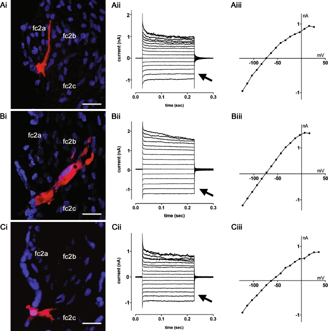
FIG. 4.
Whole-cell currents recorded from uncoupled turn 2 root cells. A–C show representative data from root cells in lateral wall slices pre-incubated with 2 mM 1-octanol. Ai Neurobiotin labeling (red) revealed a single root cell possessing a process extending between the type 2a fibrocytes (fc2a) of the spiral prominence. Nuclei were stained with DAPI (blue). Aii The root cell displayed weakly rectifying whole-cell currents in response to voltage steps (−124 mV to +26 mV, −74 mV holding potential). Aiii Current–voltage (I–V) relationship for traces shown in Aii. Bi Neurobiotin labeling of a root cell with a process extending between the type 2b fibrocytes (fc2b). Bii The root cell displayed weakly rectifying currents (−124 mV to +16 mV, −74 mV holding potential). BiiiI–V relationship for traces shown in Bii. Ci Neurobiotin labeling of a root cell with a bifurcating process extending towards type 2c fibrocytes (fc2c). Cii This root cell also displayed weakly rectifying currents (−124 mV to +36 mV, −64 mV holding potential). CiiiI–V relationship for traces shown in Cii. The arrows in Aii, Bii, and Cii denote the comparable inward currents at hyperpolarized potentials in the three cells. Scale bars = 20 µm.

