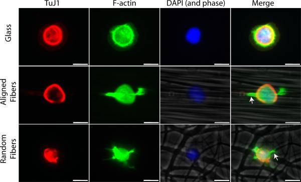
Figure 9.
Lamellipodia formation is spatially restricted on fibers. Motor neurons were grown on glass, aligned fibers, and random fibers and fixed after 1.5 h in culture. Cells were stained with Phalloidin (green), TuJ1 (red), and DAPI (blue) to visual lamellipodia. Aligned and random fibers were imaged in phase-contrast and included in the merged image. Flattened, veil-like lamellipodia were observed on glass while lamellipodia typically formed only along fibers just adjacent to the cell body (arrows). Scale bar=10 μm.

