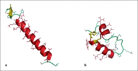
Fig. 1.
a Molecular graphics illustration of SP-C33 in methanol based on the structure of native porcine SP-C (1SPF). Helical residues in the transmembrane sequence are in red, the N-terminal serine residues are in yellow, and disordered segments are in green. b Mini-B structure in methanol based on the structure of Mini-B (1SSZ) with the helical segments in red, the engineered loop in green and the disulfide linkages in yellow.

