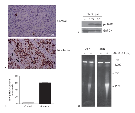
Fig. 3.
Irinotecan treatment induced DNA damage in tumor cell nuclei of FaDu xenografts. a FaDu tumor xenografts were removed 24 h after the second dose of irinotecan, were fixed and processed for the immunohistochemical staining with anti-p-H2AX. Photomicrographs are representative of the whole tumor section. In the upper panel the control has only a few tumor cell nuclei positive for p-H2AX, whereas in the lower panel the irinotecan-treated tumor show high expression of p-H2AX protein in many tumor cell nuclei. Arrows show the absence of p-H2AX in the stromal cell nuclei. b H2AX-positive tumor cells were counted and the percentage was calculated and presented in the chart showing the increase with irinotecan treatment. c p-H2AX protein levels as detected by Western blot analysis increased significantly in FaDu cells treated with SN-38. GAPDH was used as loading control. d Double-strand DNA breaks at 24 and 48 h after SN-38 (0.1 μM) treatment as observed by pulsed-field gel electrophoresis in FaDu cells. A megabase DNA damage was observed, as shown at 1,900 kb size.

