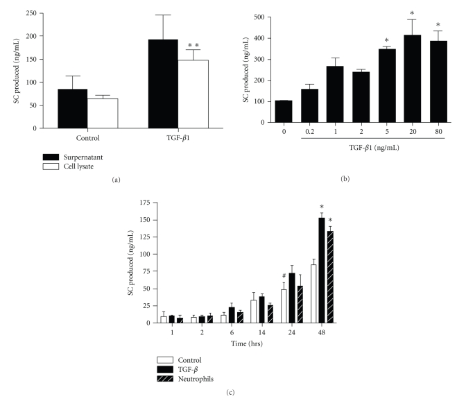
Figure 4.
Effect of TGF-β1 on pIgR/SC production. (a) Calu-3 cells were cultured for 48 hrs with or without TGF-β1 (20 ng/ml). SC production was evaluated in supernatants (solid bars) and in cell lysates (open bars). (b) Calu-3 cells were exposed to increasing concentration of TGF-β1 (0.2 to 80 ng/ml) for 48 hrs, and SC was measured in supernatants. (c) SC production was assessed in supernatants from resting Calu-3 cells as control (open bars), Calu-3 cells incubated with TGF-β1 (20 ng/ml, solid bars) and Calu-3 cells/neutrophil cocultures (stripped bars) for 1 hr to 48 hrs. Results are expressed as means ± SEM for (a) (n = 7 independent experiments with triplicates), (b) (n = 3) and (c) (n = 3). *P < .05 as compared with control (a, b, c); #P < .05 as compared with control at 1 hr (c).

