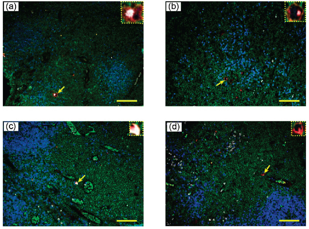
Figure 5.
Multiplexed QD staining images of lymph node biopsies from two patients with “suspicious” lymphoma (a and b), and from two patients with reactive lymph nodes (c and d). The expanded insets reveal the presence of rare malignant HRS cells in (a) and (b) but the absence of such cells in the biopsies of reactive lymph node patients (c and d). Scale bar: 100 µm.

